43 images of compound microscope with labels
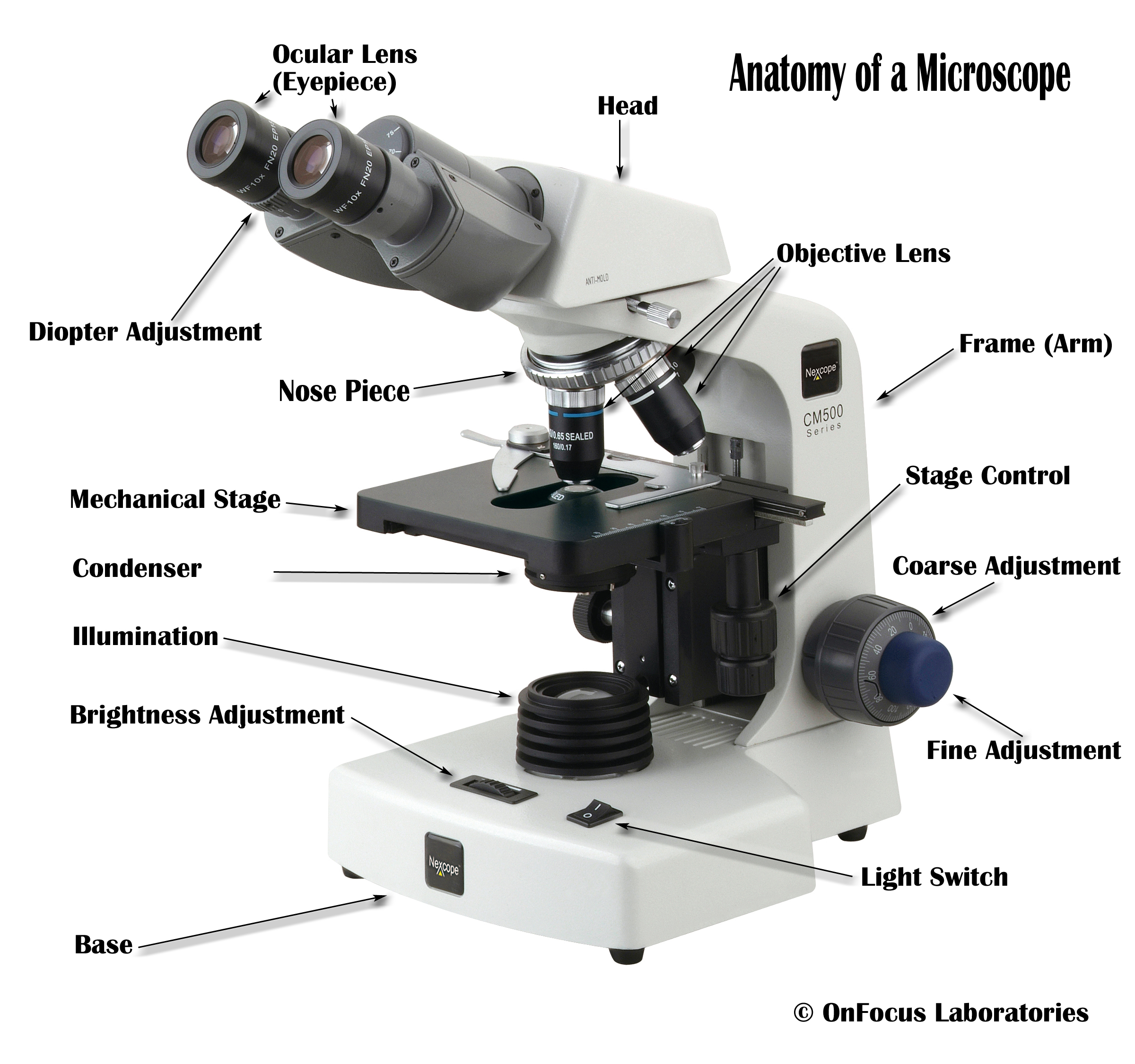
Compound Microscope Parts, Functions, and Labeled Diagram Compound Microscope Definitions for Labels. Eyepiece (ocular lens) with or without Pointer: The part that is looked through at the top of the compound microscope. Eyepieces typically have a magnification between 5x & 30x. Monocular or Binocular Head: Structural support that holds & connects the eyepieces to the objective lenses. Compound Microscope with labels Stock Vector | Adobe Stock Download Compound Microscope with labels Stock Vector and explore similar vectors at Adobe Stock. Adobe Stock. Photos Illustrations Vectors Videos Audio Templates Free Premium Editorial Fonts. ... Get 10 free Adobe Stock images. Start now. Get 10 free images. Unlock 200M+ assets in our full collection.
Compound Microscope Stock Photos and Images - Alamy Find the perfect compound microscope stock photo. Huge collection, amazing choice, 100+ million high quality, affordable RF and RM images. ... Method of illuminating compound microscope with gas lamp. Labels: C, ... Full-size copy of Robert Hooke's Compound Microscope - held at the Science Museum, London. Hooke, ...
Images of compound microscope with labels
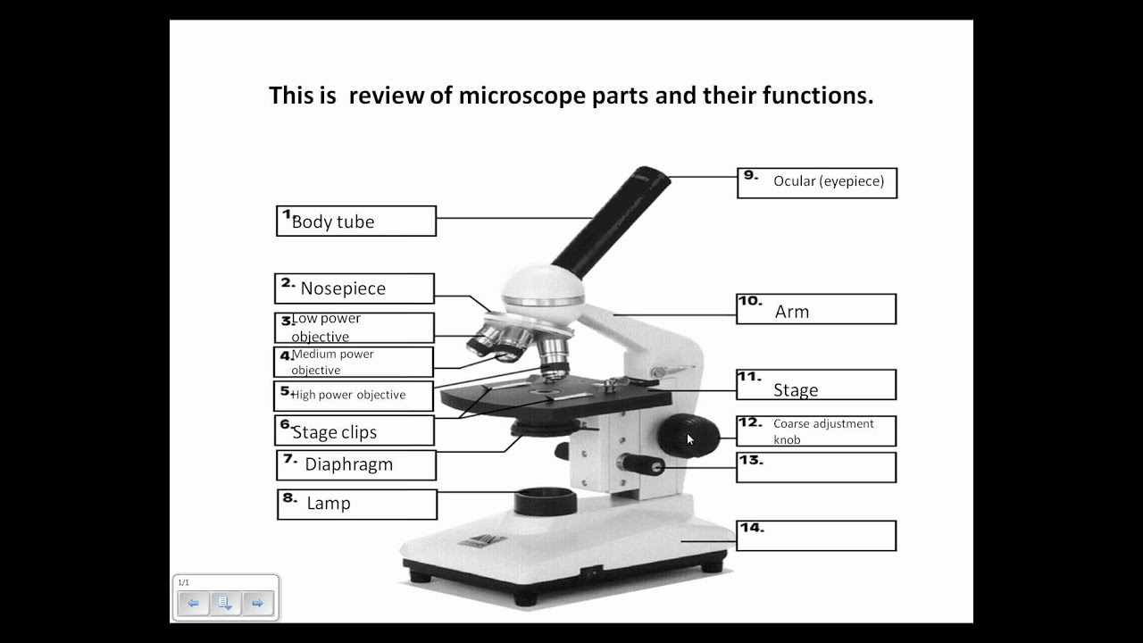
18,701 Microscope drawing Images, Stock Photos & Vectors - Shutterstock Microscope drawing royalty-free images 18,701 microscope drawing stock photos, vectors, and illustrations are available royalty-free. See microscope drawing stock video clips Image type Orientation Color People Artists Sort by Popular Science Abstract Designs and Shapes College and University Art Styles Printing, Typography, and Calligraphy microscope picture with labels - Compound Light Microscope... View microscope picture with labels from BIOL 1005Y at Yeshiva University. Compound Light Microscope ocular (eyepiece) revolving nosepiece objectives coarse adjustment knob mechanical stage fine Compound Microscope - Types, Parts, Diagram, Functions and Uses It comes with a wide body and base. Its distinct parts include a condenser, illumination, focus lock, mechanical stage, and a revolving nosepiece which can hold up to five objectives. It usually has a binocular head, which makes long-term observation easy. Image 22: An example of a research compound microscope.
Images of compound microscope with labels. Compound microscope with label and picture? - Answers Picture and Image Searches Create. 0. Log in. Compound microscope with label and picture? Wiki User. ∙ 2012-06-19 11:30:43. Study now. See answer (1) Best Answer. Copy. yes85235252. Parts of a Compound Microscope - Labeled (with diagrams) A compound microscope is known as a high-power microscope that enables you to achieve a high level of magnification. Smaller specimens can be thoroughly viewed using a compound microscope. ... Image 3: A compound microscope with a corresponding label of the different parts. imagesource: images.slideplayer.com ... Labels: microsopes Newer Post ... A Study of the Microscope and its Functions With a Labeled Diagram To better understand the structure and function of a microscope, we need to take a look at the labeled microscope diagrams of the compound and electron microscope. These diagrams clearly explain the functioning of the microscopes along with their respective parts. Man's curiosity has led to great inventions. The microscope is one of them. Parts of a microscope with functions and labeled diagram - Microbe Notes The higher the magnification of the condenser, the more the image clarity. More sophisticated microscopes come with an Abbe condenser that has a high magnification of about 1000X. Diaphragm - it's also known as the iris. Its found under the stage of the microscope and its primary role is to control the amount of light that reaches the specimen.
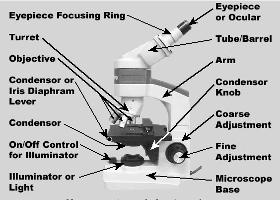
Compound microscope Images, Stock Photos & Vectors | Shutterstock Compound microscope images 3,117 compound microscope stock photos, vectors, and illustrations are available royalty-free. See compound microscope stock video clips Image type Orientation Sort by Popular Science College and University Biology Insects and Spiders Jobs/Professions microscope laboratory compound eye optical microscope scientist Next Compound Microscope - Diagram (Parts labelled), Principle and Uses See: Labeled Diagram showing differences between compound and simple microscope parts Structural Components The three structural components include 1. Head This is the upper part of the microscope that houses the optical parts 2. Arm This part connects the head with the base and provides stability to the microscope. › AMSCOPE-KIDS-Prepared-MicroscopeAMSCOPE-Kids 48pcs Kids Plastic Prepared Microscope Slides of ... 48 kids microscope prepared slides of entomology, botany and mammology specimens for kids, entry-level students or home school programs ; Selected fine specimens from various categories, colored labels enable an easy classification ; Dimensions: 60mm x 20mm x 1 mm. Material: plastics ; Brand new from factory. Microscope Parts and Functions First, the purpose of a microscope is to magnify a small object or to magnify the fine details of a larger object in order to examine minute specimens that cannot be seen by the naked eye. Here are the important compound microscope parts... Eyepiece: The lens the viewer looks through to see the specimen.
› zh_en › p104268Cas(9003-01-4), Poly(acrylic acid) 50% solution-Aladdin, PAA, Poly (acrylic acid) (PAA) is hygroscopic, brittle and colorless in nature with Tg at nearly 106oC. At temperatures above 200 to 250oC, it loses water and becomes an insoluble crosslinked polymer anhydride. Labeling the Parts of the Microscope | Microscope World Resources Labeling the Parts of the Microscope This activity has been designed for use in homes and schools. Each microscope layout (both blank and the version with answers) are available as PDF downloads. You can view a more in-depth review of each part of the microscope here. Download the Label the Parts of the Microscope PDF printable version here. Microscope With Labels clip art | Microscope parts, Scientific method ... Jul 3, 2012 - Download Clker's Microscope With Labels clip art and related images now. Multiple sizes and related images are all free on Clker.com. en.wikipedia.org › wiki › Electron_microscopeElectron microscope - Wikipedia An electron microscope is a microscope that uses a beam of accelerated electrons as a source of illumination. As the wavelength of an electron can be up to 100,000 times shorter than that of visible light photons , electron microscopes have a higher resolving power than light microscopes and can reveal the structure of smaller objects.
Microscope Diagram to Print
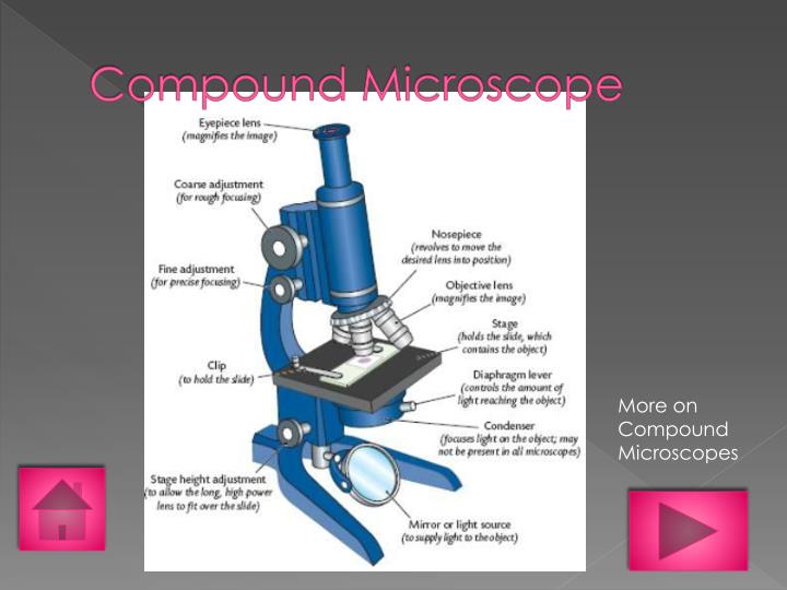
Compound Microscope: Definition, Diagram, Parts, Uses, Working ... - BYJUS A microscope with a high resolution and uses two sets of lenses providing a 2-dimensional image of the sample. The term compound refers to the usage of more than one lens in the microscope. Also, the compound microscope is one of the types of optical microscopes. The other type of optical microscope is a simple microscope.

Microscope Lab: The Compound Microscope by Amy Brown Science | TpT
Compound Microscope- Definition, Labeled Diagram, Principle, Parts, Uses In order to ascertain the total magnification when viewing an image with a compound light microscope, take the power of the objective lens which is at 4x, 10x or 40x and multiply it by the power of the eyepiece which is typically 10x. Therefore, a 10x eyepiece used with a 40X objective lens will produce a magnification of 400X.
1.Fully label the compound microscope below.
› seterra › en-anMicroscope Components - Science Quiz - GeoGuessr Microscope Components - Science Quiz: The most common type of modern microscope is called a compound microscope. They have two systems of lenses, one is the eyepiece and the other is comprised of one or more objective lenses. This type of microscope has become so advanced that some are capable of magnifying up to 1000 times! Microscopes are used in almost all types of scientific research, and ...

BIOLOGY BASICS - "Compound Microscope: Basic Use" - YouTube
Compound Microscope Labeled Diagram | Quizlet QUESTION. The total magnification of a specimen being viewed with a 10X ocular lens and a 40X objective lens is. 15 answers. QUESTION. a mosquito beats its wings up and down 600 times per second, which you hear as a very annoying 600 Hz sound. if the air outside is 20 C, how far would a sound wave travel between wing beats. 2 answers.

Compound Microscope, कंपाउंड माइक्रोस्कोप in Sector 24 , Uniteck Scientific & Electronic ...
› books › NBK26880Looking at the Structure of Cells in the Microscope ... A typical animal cell is 10–20 μm in diameter, which is about one-fifth the size of the smallest particle visible to the naked eye. It was not until good light microscopes became available in the early part of the nineteenth century that all plant and animal tissues were discovered to be aggregates of individual cells.

How to Use a Compound Microscope: 11 Steps (with Pictures)
10 Best Compound Microscopes (Summer 2022) - The Complete Guide Celestron Labs designed a premium compound microscope with exceptional durability. It comes with a two-year warranty, but it will last longer if you maintain it right. The product includes two eyepieces, a WP-20x and a WF-10x with a pointer. You can change these eyepieces easily to find your perfect adjustment.
23 Label And Color The Parts Of Both Microscopes - Labels 2021
Labeled microscope diagram - Pinterest A diagram showing all of the parts of a compound light microscope. Racing is really fun, especially if you can express your creativity through creating your own car. Here are the awesome pinewood derby templates that will inspire you and help you build a cool car easily. Art, Humor, & More!
PPT - Types of Microscopes PowerPoint Presentation - ID:1588009
Compound Microscope Parts - Labeled Diagram and their Functions Basically, compound microscopes generate magnified images through an aligned pair of the objective lens and the ocular lens. In contrast, "simple microscopes" have only one convex lens and function more like glass magnifiers. [In this figure] Two "antique" microscopes played significant roles in the history of biology.
Microscope Review.wmv - YouTube
› cemf › whatisemWhat is Electron Microscopy? - UMASS Medical School Conventional scanning electron microscopy depends on the emission of secondary electrons from the surface of a specimen. Because of its great depth of focus, a scanning electron microscope is the EM analog of a stereo light microscope. It provides detailed images of the surfaces of cells and whole organisms that are not possible by TEM.

All Saints Online: Microscope Part Functions
Simple Microscope - Diagram (Parts labelled), Principle, Formula and Uses The working principle of a simple microscope is that when a lens is held close to the eye, a virtual, magnified and erect image of a specimen is formed at the least possible distance from which a human eye can discern objects clearly. Magnification formula The magnification power of a simple microscope is expressed as: M = 1 + D/F Where

Anatomy and Physiology I Coursework: Microscope A+P
Picture Of Microscope With Label, Compound Light Microscope Labeled ... Although there are now many advanced microscope types, some applications may still demand the use of a simple microscope. It consists of simple parts and performs simple functions. Where the slide is held/placed. Label free imaging possible with this ultimate three dimensional. Photo compound microscope with labels image 3850568.

Anatomy and Physiology I Coursework: Microscope A+P
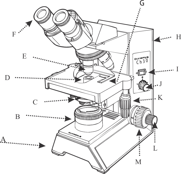
Solved Label the image of a compound light microscope using - Chegg Expert Answer. 100% (17 ratings) Transcribed image text: Label the image of a compound light microscope using the terms provided.

Anatomy and Physiology I Coursework: Microscope A+P
Labelled Diagram of Compound Microscope - Biology Discussion The below mentioned article provides a labelled diagram of compound microscope. Part # 1. The Stand: The stand is made up of a heavy foot which carries a curved inclinable limb or arm bearing the body tube. The foot is generally horse shoe-shaped structure (Fig. 2) which rests on table top or any other surface on which the microscope in kept.

34 Label Microscope Quiz - Labels Information List
16 Parts of a Compound Microscope: Diagrams and Video In compound microscopes with two eye pieces there are prisms contained in the body that will also split the beam of light to enable you to view the image through both eye pieces. 2. Arm. The arm of the microscope is another structural piece. The arm connects the base of the microscope to the head/body of the microscope.
Compound Microscope
Parts of the Microscope with Labeling (also Free Printouts) Parts of the Microscope with Labeling (also Free Printouts) By Editorial Team March 7, 2022 A microscope is one of the invaluable tools in the laboratory setting. It is used to observe things that cannot be seen by the naked eye. Table of Contents 1. Eyepiece 2. Body tube/Head 3. Turret/Nose piece 4. Objective lenses 5. Knobs (fine and coarse) 6.

CHAPTER 5: THE FUNDAMENTAL UNIT OF LIFE - NCERT Class 9 Science For Blind and Visually Impaired ...
› publication › 320945390(PDF) Introduction to Microscopy - ResearchGate Nov 08, 2017 · • In compound microscope it will be i.e 10 X, f= 16 mm; 40 X, f= 4 mm; 100 X, f= 1.8 mm. • Image produced by objective lens falls on the eyepiece lens serve as objec t. • Image formed in the ...
Post a Comment for "43 images of compound microscope with labels"